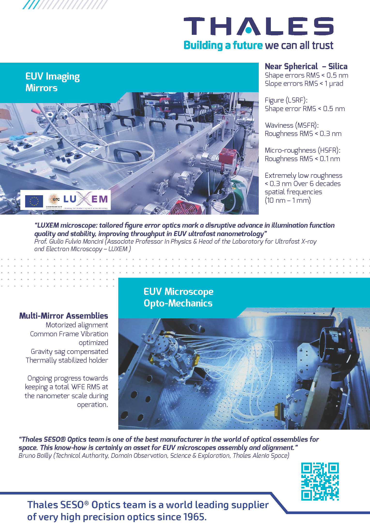
2025-11-17T22:28:46+00:00

“Explore the behind-the-scenes of scientific successes by photographing those who build these successes” – ERN 2024 In September 2024, photographs of LUXEM Team and Research Facilities taken by the “Associazione Fotografica Città Giardino” were highlighted in the European Researchers Night exhibit at the Broletto municipality palace in Pavia. Find out more

2025-11-17T22:28:46+00:00

2024-03-27T09:59:00+00:00

Cover – OPTICA Vol. 11, Issue 3, pp. 403-411 Twisted-phase OAM illumination and its resulting diffraction pattern in single-shot ptychography experiments at an FEL source. Find out more

2024-03-27T09:59:00+00:00

Il Sole24Ore – 20th December 2023

2025-11-17T22:21:29+00:00

Il Sole 24 Ore LUXEM projects ERC StG ULTRAIMAGE and ERC PoC HYPER highlighted in the Italian newspaper "Il Sole24Ore". In the article appeared on Wednesday 20th December 2023, on "Il Sole24Ore", Prof. Mancini explains relevance and breakthrough impact of the 2 ERC projects @LUXEM. Find out more

Il Sole24Ore – 20th December 20232025-11-17T22:21:29+00:00

Women in Science Winners of Competitive Cariplo Foundation Calls

2025-11-17T22:31:58+00:00

Women in Science Winners of Competitive Cariplo Foundation Calls Prof. Mancini’s and LUXEM Research highlighted in two Stories from Cariplo Foundation: “Women in Science – Cariplo Researchers” and “Winners of Cariplo Research Grants” Women in Science - Cariplo Researchers Winners of Cariplo Research Grants

Women in Science Winners of Competitive Cariplo Foundation Calls2025-11-17T22:31:58+00:00

Fast-moving plot

2025-11-17T22:33:24+00:00

Fast-moving plot How do dye-sensitised solar cells work, and what's behind the brilliant new mobile phone displays? The ultrashort X-ray pulses at SwissFEL reveal the chemical reactions that take place inside these devices and could help to make them even more efficient and cost-effective. Find out more

Fast-moving plot2025-11-17T22:33:24+00:00

Ptychography – With a “P” As in “Pterodactyl”

2022-11-14T19:41:54+00:00

Ptychography – With a “P” As in “Pterodactyl” In this invited blog from DECTRIS Dr. Charles Bevis and Prof. Giulia Mancini discuss the development of Ptychographic Coherent Diffractive Imaging and the possibilities it offers. What is ptychography? How does it work? Is it worth all the hype? Find out more

Ptychography – With a “P” As in “Pterodactyl”2022-11-14T19:41:54+00:00

Cover – The Journal of Physical Chemistry Letters

2022-05-01T21:48:14+00:00

Cover – The Journal of Physical Chemistry Letters Volume 13, Issue 14, pages 3310-3528 We provide a comprehensive atomic-scale description of the purely thermal response of lead-halide perovskites in a temperature range relevant for realistic applications. Thermal-induced changes are rationalized in terms of displacive phase transitions, which are a consequence of the phonon anharmonicity of the inorganic lattice. Find out more

Cover – The Journal of Physical Chemistry Letters2022-05-01T21:48:14+00:00

ACS Editors’ Choice

2023-07-03T09:23:47+00:00

ACS Editors’ Choice Nano Letters - 2016 The design and characterization of functionalized nanoparticle supracrystals require atomically resolved information on both the metallic core and the external organic ligand shell. Ultrafast small-angle electron diffraction has enabled to resolve both static ordering properties and photo-induced motions of supracrystals of alkanethiol-capped gold nanoparticles, with combined fs temporal and Å spatial resolution. Key to this advance was the unique capability to retrieve the local symmetry of the real-space objects creating an electron speckle pattern by computing angular cross-correlation functions. This method, well known in the X-rays community, has been applied to the analysis of [...]

ACS Editors’ Choice2023-07-03T09:23:47+00:00

An innovative device studies gold nanoparticles in depth

2022-02-17T12:17:55+00:00

An innovative device studies gold nanoparticles in depth EPFL NEWS - 2016 Ultrafast Electron Diffraction exploits the high sensitivity that electrons have for interacting with matter. A unique UED device developed by Giulia Mancini and Fabrizio Carbone (EPFL), in collaboration with Jom Luiten (TU/e) and Tatiana Latychevskaia (PSI), can study monolayers and very thin samples containing light elements, e.g. hydrogen and carbon. And when it comes to dense aggregates and small molecules, the small-angle time-resolved electron diffractometer can achieve the extreme sensitivity of a traditional Free Electron Laser, but at a fraction of the cost. This set-up is a reference for [...]

An innovative device studies gold nanoparticles in depth2022-02-17T12:17:55+00:00

The Sharpest Images

2022-02-17T12:16:25+00:00

The Sharpest Images JILA News – Laser Physics Nanoscience 2017 The world’s most powerful CDI (Coherent Diffractive Imaging) microscope at a wavelength of 13 nm­­. Remarkably, it contains no lenses. Rather, a nanoscale object is illuminated with coherent high-harmonic beams of extreme ultraviolet light. The scattered light is collected and used with a powerful algorithm to create an image of the object with sub-wavelength spatial resolution. Find out more

The Sharpest Images2022-02-17T12:16:25+00:00
Go to Top