LUXEM Facilities
The ability to visualize in real time charge and energy transport in materials interacting with light pulses is a major current challenge in nanotechnology, relevant to a host of different applications, including photovoltaics and optoelectronic devices. The design of such materials relies critically on the availability of new, accurate, reliable, and fast characterization tools capable of quantifying how materials’ functionality and light-induced performance are related to their electronic and structural properties at the nanoscale.
The Ultrafast X-ray and Electron Microscopy (LUXEM) laboratory addresses this challenge by developing new, cutting-edge ultrafast microscopy devices, which it uses to understand the response of materials to ultrashort light pulses during the very first moments of radiation-matter interaction, with resolutions down to a few millionths of a meter in space and a few millionths of a billionth of a second in time. We study structure-property relationships in novel nanomaterials from hierarchical nanoparticles assembly, watching transport processes occur at interfaces in real-time. We do so by developing and implementing innovative methods for ultrafast in-situ and in-operando microscopy characterized by femtosecond (10-15s) time- and Ångstrom-to-nanometer spatial resolution. These high-tech approaches integrate tabletop pulsed EUV/X-ray photons from High-Harmonic Generation (HHG) and electrons. We collaborate with Synchrotron and Free-Electron Laser large-scale facilities.
LUXEM’s facilities include two laboratories:
- LUXEM 1: ultrafast laser laboratory
- LUXEM 2: prototyping, metrology, and educational laboratory
LUXEM 1
The laboratory is located in a newly renovated infrastructure at the Department of Physics at the University of Pavia (IT). It features a stabilized temperature and humidity environment with vibration isolation, an independent thermal regulation unit, and back-up generator. The Laboratory hosts:
- A Ti:sapphire ultrafast regenerative amplifier that employs a patented cryogenically-cooled amplifier technology with tunable average pulse energy
- A tabletop time-resolved microscopy and nanometrology beamline with EUV/soft X-ray light from High-Harmonic Generation, where we obtain images from diffraction patterns through computational iterative algorithms.
- A compact beamline for ultrafast electron diffraction and small-angle scattering, where we also develop new methods of image reconstruction and speckle detection from compact sources with partial spatial coherence
The beamlines are fully custom designed within LUXEM, in collaboration with international academic and industrial partners. They both are implemented in ultra-high vacuum [10-7 to 10-10mbar] on a highly-stabilized vibration damped optical table, which also hosts the ultrafast laser source. The set-ups are designed to run in parallel at 5kHz or, separately, at higher rep-rates. Both beamlines feature capabilities for experiments in transmission or reflection geometry with high samples throughput and full remotely controlled actuation systems. Their operating configurations are selected depending on the signal-to-noise, flux, jitter, dose, required for each experimental campaign.

The ultrafast laser source
A Ti:sapphire ultrafast regenerative amplifier that employs a patented cryogenically-cooled amplifier technology with tunable average pulse energy: > 3 mJ at 5 kHz, > 2 mJ at 10 kHz, > 0.6 mJ at 20 kHz, with 35fs average pulse duration and 785nm center wavelength (KMLabs). The laser system is equipped with a pulse-shaper and a stabilization system for long-term beam drifts. This source feeds two beamlines for: (i) ultrafast soft X-ray spectro-microscopy and (ii) ultrafast electron diffraction imaging. In each beamline, the amplified femtosecond laser beam is divided into two paths:
- The pump laser pulse, which initiates the sample’s dynamics and serves as a reference point in time
- The probe – in our case extreme ultraviolet/soft x-ray photons or electrons pulses, which record image snapshots of the samples’ dynamics as a function of the time delay
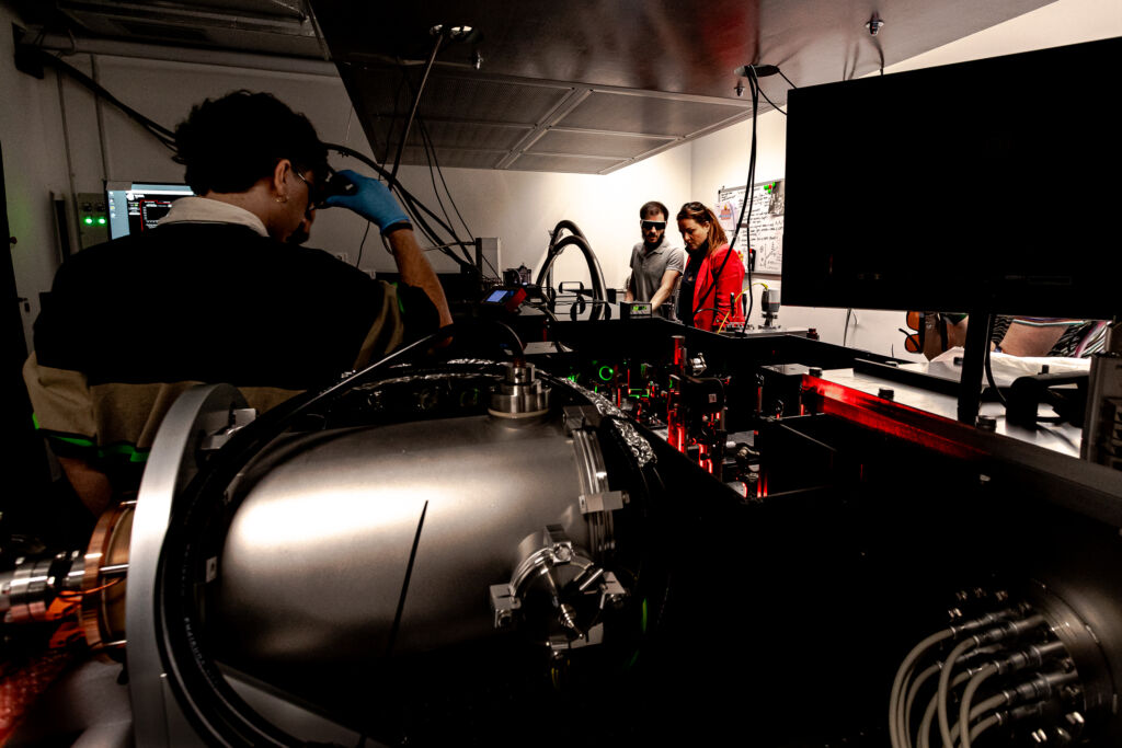
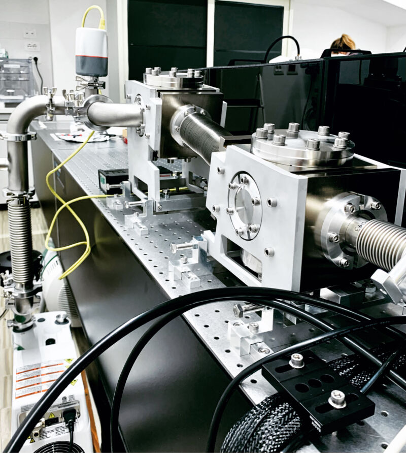
The Ultrafast EUV/soft X-ray Microscopy beamline
Ultrafast EUV microscopy is primarily carried-out (albeit not exclusively) with a technique for coherent diffractive imaging called ptychography, in which multiple diffraction patterns from overlapping fields of view are processed by iterative algorithms to recover amplitude and phase images of sample and beam, separately.
The probe, i.e. EUV/soft X-ray light pulses, is generated via a non-linear process called High-Harmonic Generation (HHG). In HHG, the laser light from the tabletop femtosecond laser is coherently upconverted to shorter wavelengths while retaining excellent spatial coherence when implemented in a phase-matched geometry, as well as temporal coherence, with durations of <100 attoseconds. In our microscope, we generate bright, phase-matched beams in a hollow-core waveguide at 13nm (1010 photons/sec, He at 400-700 torr) or 30nm (1012 photons/sec, Ar at 30-60 torr). The fundamental driving frequency is eliminated before the sample with rejecter mirrors and metallic filters. The beamline is equipped with an in-line intensity monitor and a spectrometer capable of recording I0 beam data and monitoring the spectral quality of the probe beam every 100 pulses at 5kHz.
The ultrafast microscope chamber is custom-designed for full remotely controlled in-vacuum positioning of all optical elements, the sample, and the detector, allowing for a flexible range of Numerical Apertures (NA) from 0.04-0.5, or diffraction-limited spatial (transverse) resolutions from 150nm down to 13nm with 13nm illumination. The EUV light is focused and overlapped with the pump pulses nearly collinearly onto the sample; their arrival time is controlled by a motorized delay-line. At each time-delay, multiple coherent patterns from overlapping fields of view are collected on an in-vacuum EUV CMOS detector with 11µm pixel size at a maximum rate of 24 fps (AXIS Photonique), without the need for image-forming optics.
In ptychography, iterative phase-retrieval algorithms are used to reconstruct the sample phase information, lost when recording only the diffraction intensity. The dynamic response of each sample is captured by collecting stroboscopic images as a function of time delay between pump and probe pulses, acquiring amplitude and phase snapshots of a nanoscale movie.
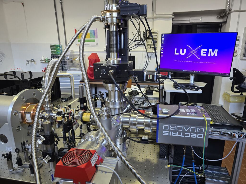
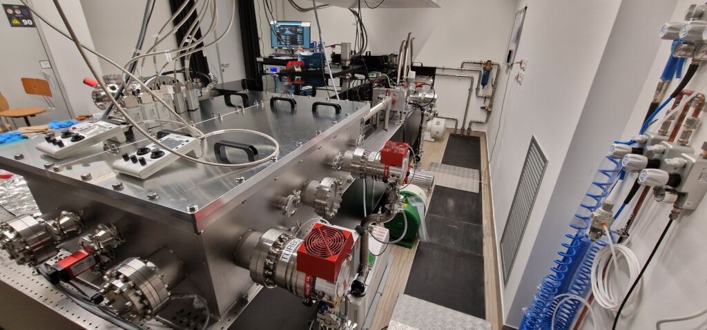
The Ultrafast Electron Diffraction Imaging beamline
In this beamline for time-resolved electron diffraction, we also develop new methods for image reconstruction and speckle detection from compact sources with partial spatial coherence. It is also possible to customize the transverse coherence of the electron beam to enable speckle detection and analysis with modern techniques of angular cross-correlation.
The amplified femtosecond pulse trains are divided into the pump (optical) and the probe (non-relativistic electron bunches) paths with a beam-splitter. The probe beam is frequency-tripled by third harmonic generation in nonlinear BBO crystals, and it is used to generate the probing electron pulses inside a high-voltage (0-100kV) DC gun. The electron probe pulses are directed to the sample with two steering plates for the orthogonal deflection of the beam, and a solenoid, to focus the beam onto the sample. The sample is contained in the experimental chamber, which is designed to have openings for the incoming probe pulses, the pump optical access, the detector and a cryogenic system that allows to explore a range of temperatures from 1.8 K up to 300 K. Additional openings are reserved for sample vision & alignment, and pressure gauges. A high-precision manipulator allows the motion of the sample along three axis (x, y, z) and one angle (the rotation around the cryostat axis). The angular rotation of the sample around its surface normal is also possible through a piezo-electric sample holder, which is in thermal contact with the cryostat.
An optical delay line allows to change the time of arrival on the sample between pump and probe pulses. The electron pulses scatter from the sample and diffraction images are detected by the Hybrid Particle Counting pixelated detector QUADRO (DECTRIS), which is capable of single-shot detection and one-to-one conversion efficiency between electrons and output counts.
LUXEM 2
LUXEM 2 is a new research space built from the synergy between the European Research Council, the Cariplo Foundation, the University of Pavia, and the LUXEM Team. The laboratory’s infrastructure renovation was completed in December 2024. LUXEM 2 fulfils the following objectives:
- Prototype and validate new technology before it is interfaced with the femtosecond pulsed laser source
- Perform metrology and inspection of optics and samples using advanced optical and atomic force microscopy techniques
- Support teaching in photonics, pulsed laser physics, and microscopy through educational and student-friendly experimental setups.
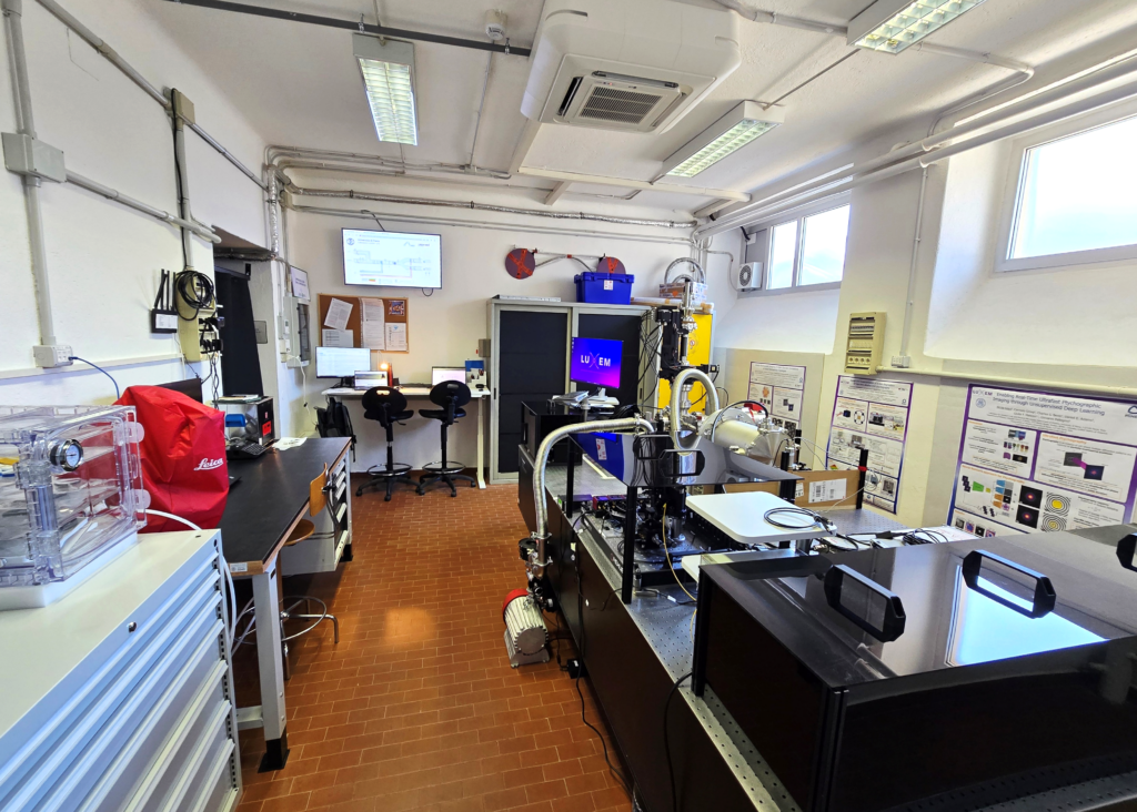
The laboratory is equipped with:
- A femtosecond titanium-sapphire oscillator (Griffin KMLabs) 80 MHz, 5 fs, 480 mW mode-locked
- A Leica DM2700 M dark and bright field optical microscope with 4 objectives and polarized light
- An Atomic Force Microscope (AFM) with:
- Scan area: 0.05 µm x 0.05 µm (Min); 20 µm x 20 µm (Max)
- Resolution: 10nm
- AFM modes: Constant Force; Constant Height; Lateral Force; Force-Distance Curves
- Educational kits for students:
- Fourier Optics
- Optical Microscopy, covering the following topics:
- Optical Imaging Basics
- Köhler Illumination
- Abbe Theory of Image Formation
- Contrast Methods
- Fluorescence Microscopy
- Spectra and Filters
- Vacuum Storage Systems for Soft X-ray Optics and Filters
- Chemical Storage Systems
Photo Gallery
LUXEM Lab – Overview
LUXEM Lab – September 2021
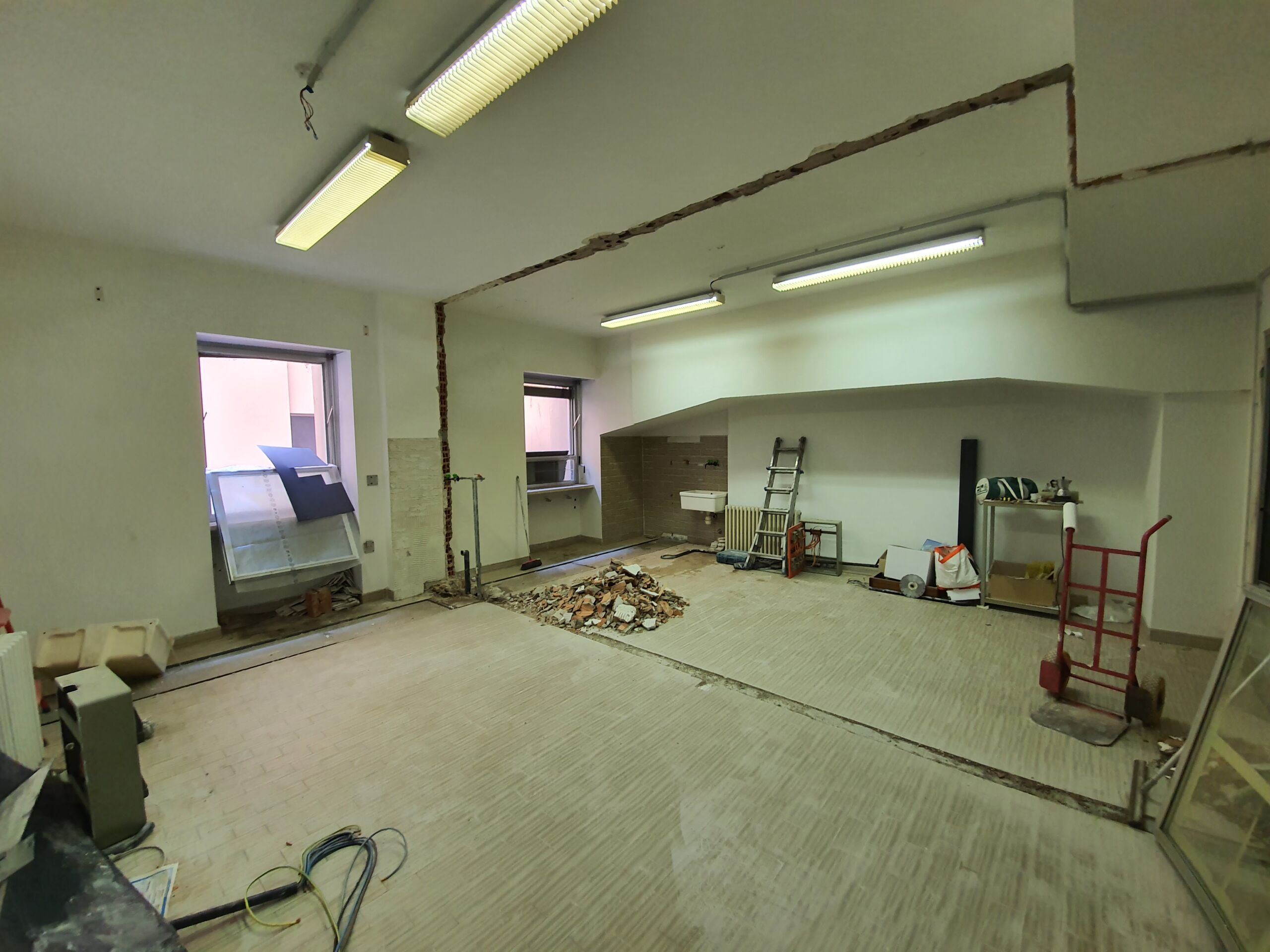
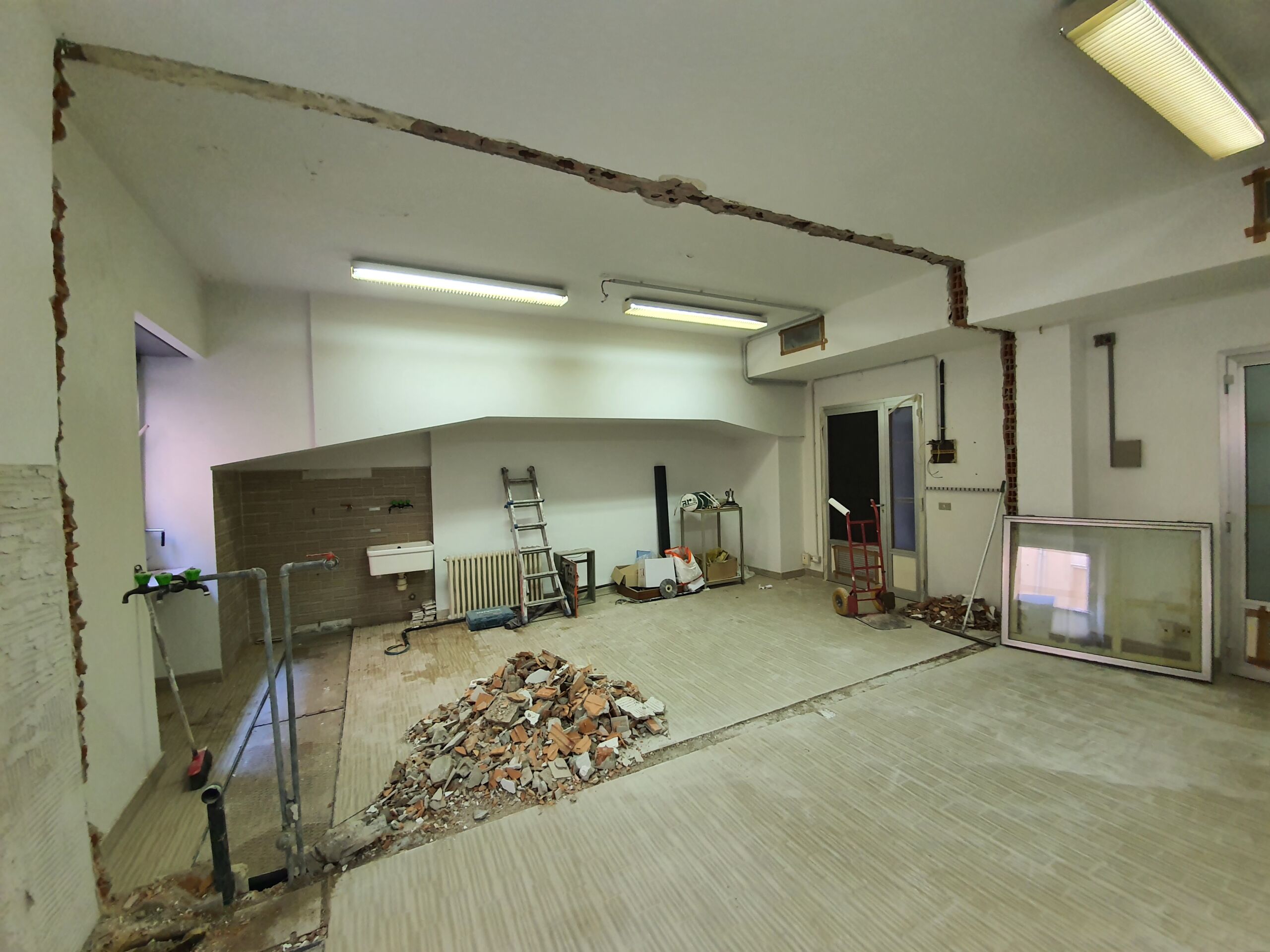
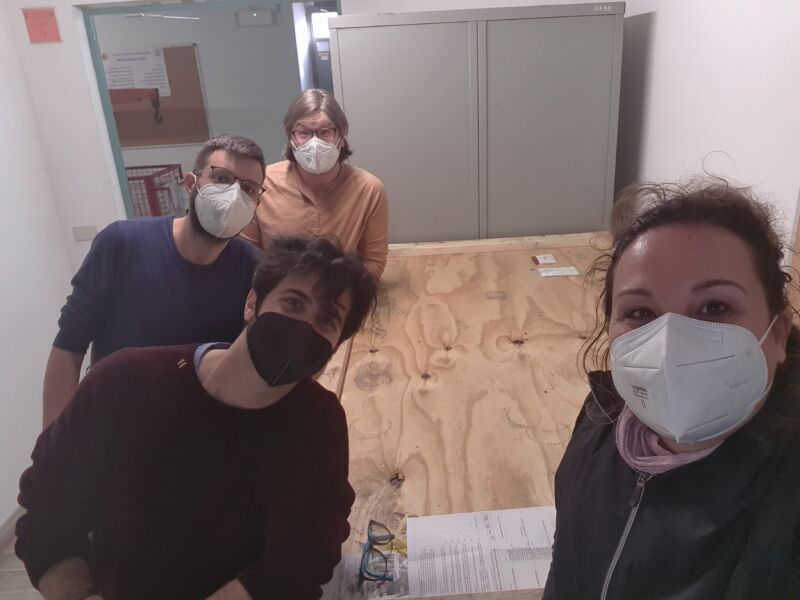
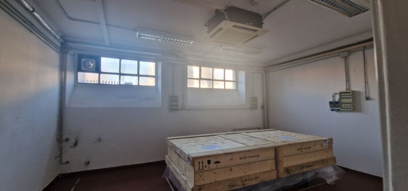
Laboratory under construction in 2021
 |
 |
 |
 |
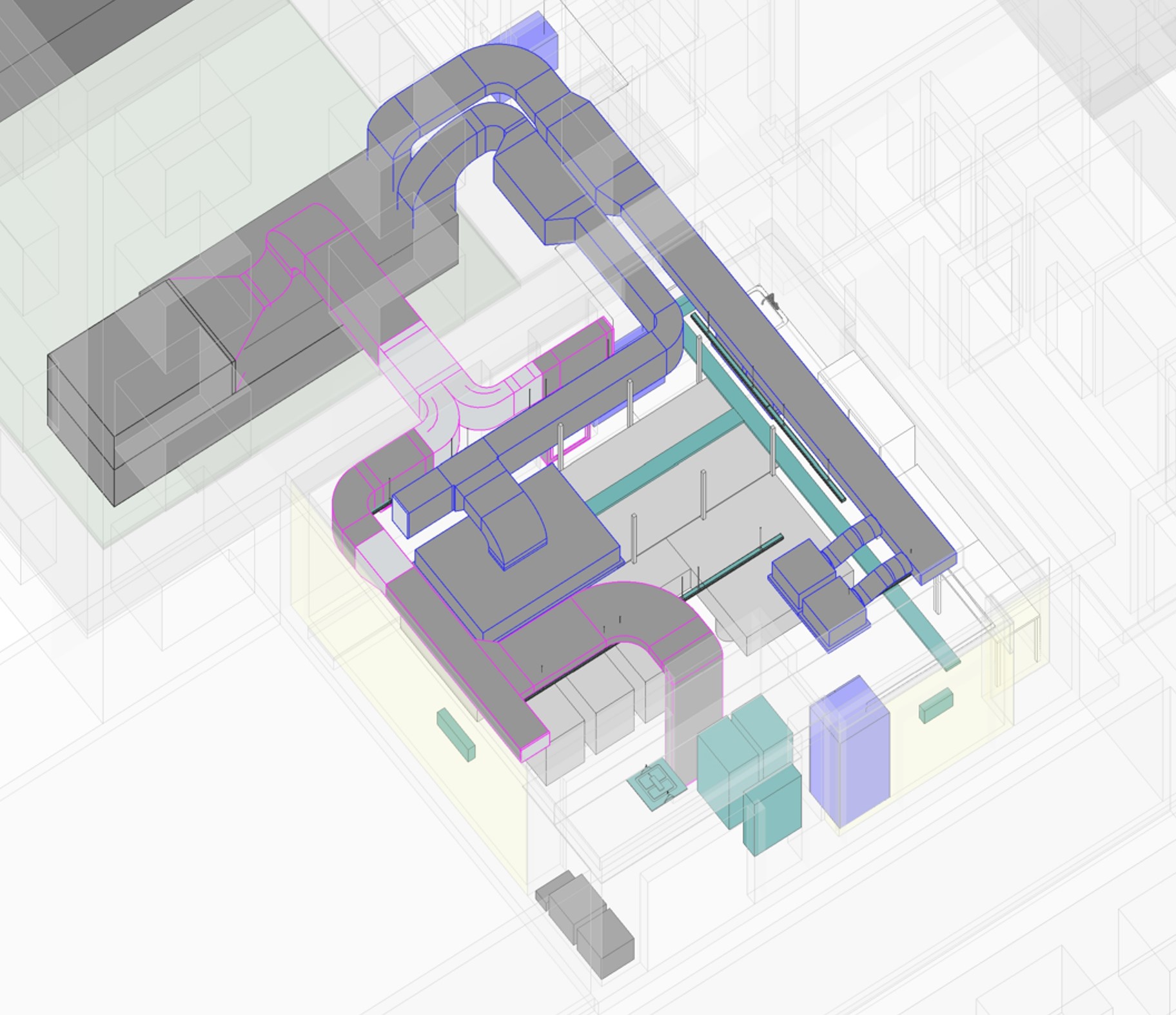
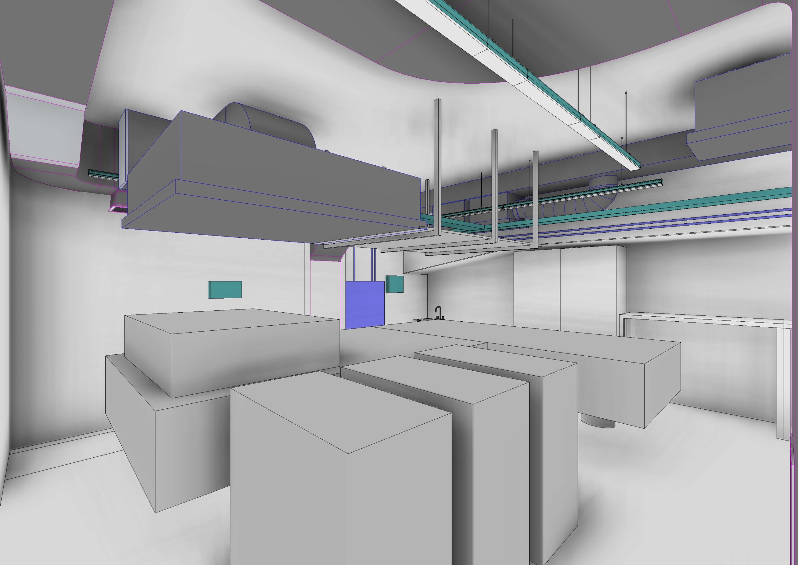
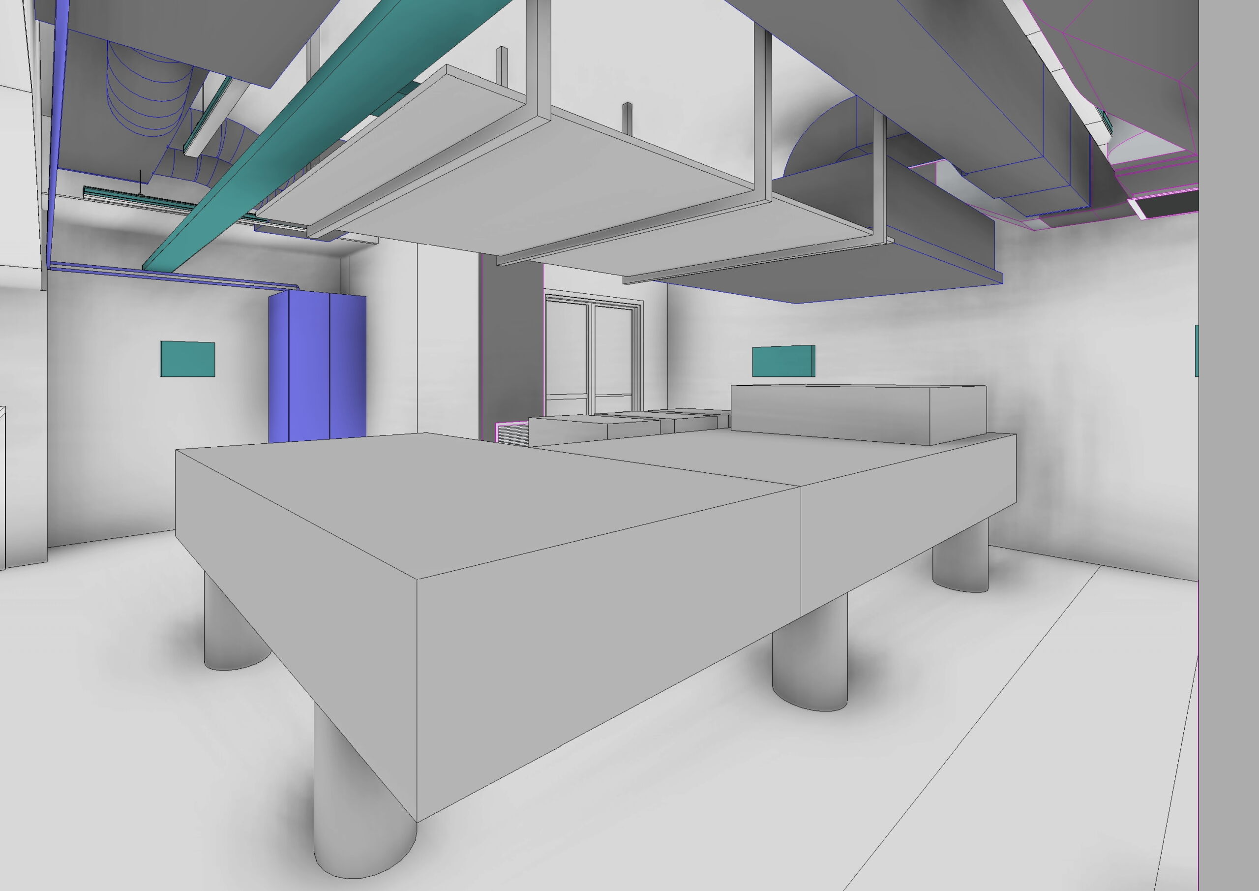
LUXEM infrastructure completion – April 2022
3D Renderings courtesy of Ing. Cristina Cecchini and Ing. Claudio Ogliari
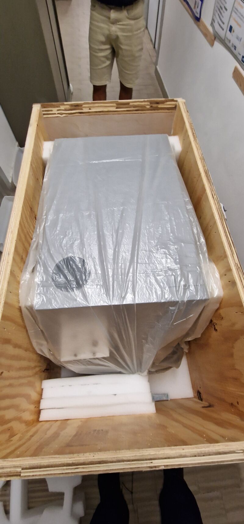
Ultrafast Laser Systems arrives to Pavia – May 2022
The RAEA Laser is moved into LUXEM
Vertical rejecters system implemented in collaboration with the INFN Mechanical Workshop and KMLabs – Mar 2023

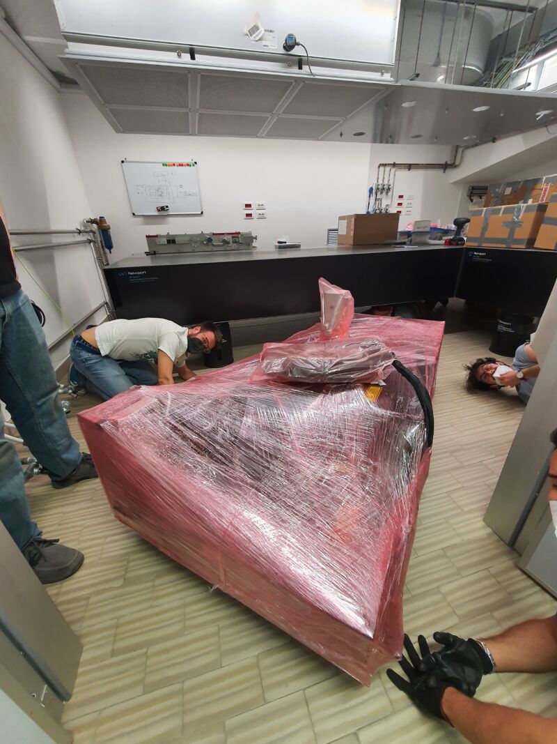
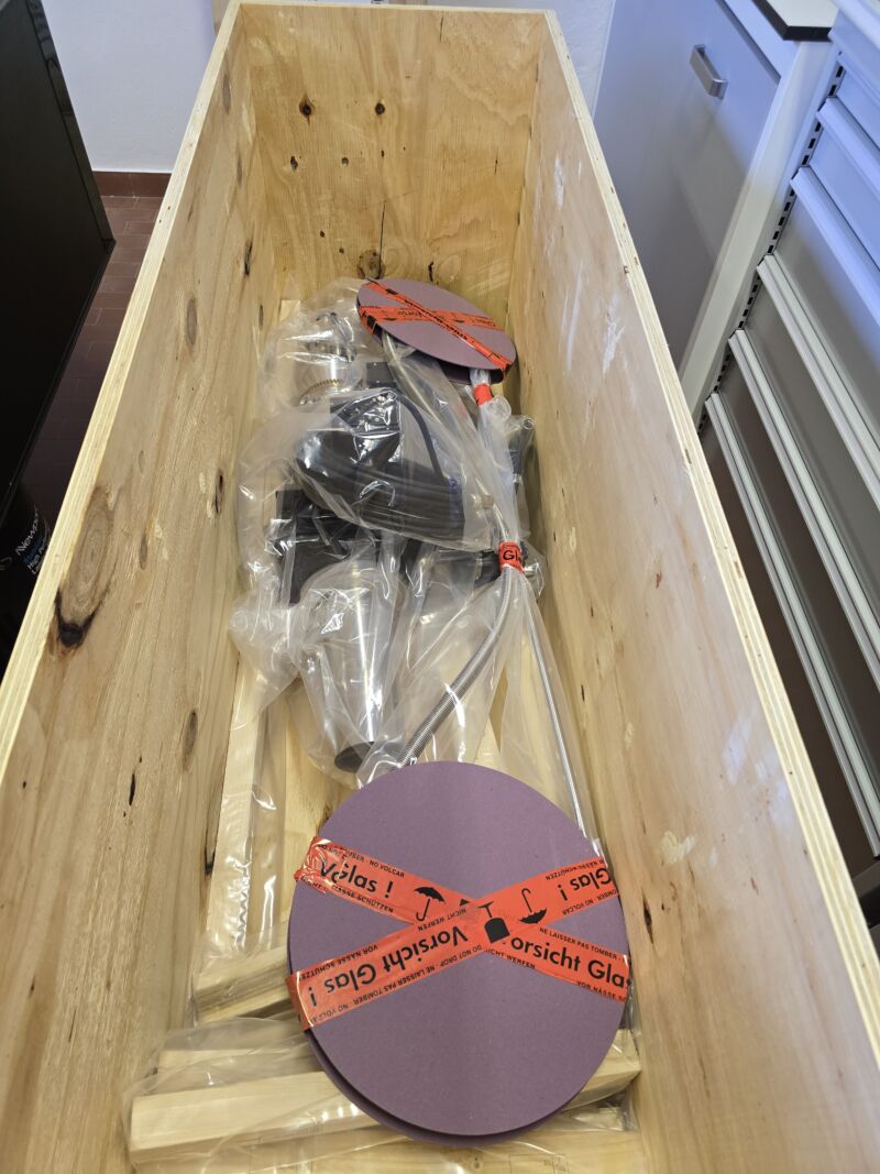
Delivery and Installation of the XUV Experimental chamber in collaboration with the INFN Mechanical Workshop – May 2023
Delivery and Installation of the XUV Experimental chamber in collaboration with the INFN Mechanical Workshop – May 2023

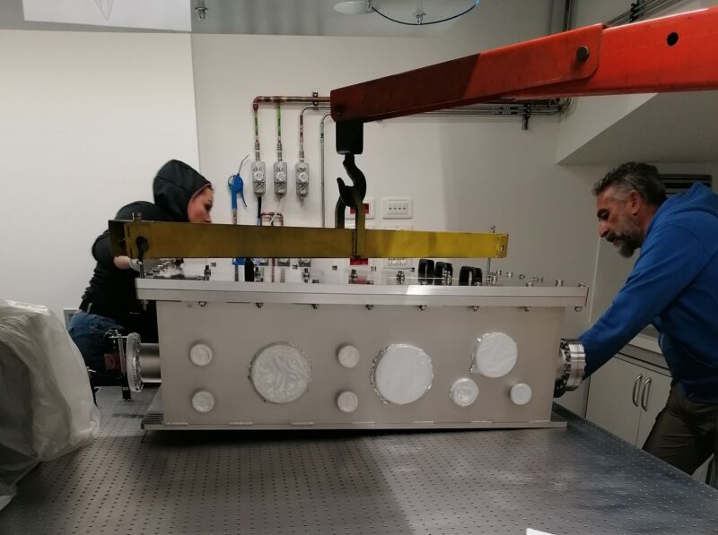
Microscope XUV experimental chamber installed at LUXEM – June 2023
Ultrafast EUV/soft X-ray Microscopy beamline first vacuum test – August 2023
Troubleshooting of the Laser pump main chiller – September 2023
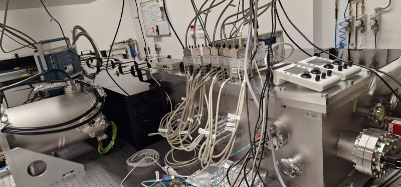
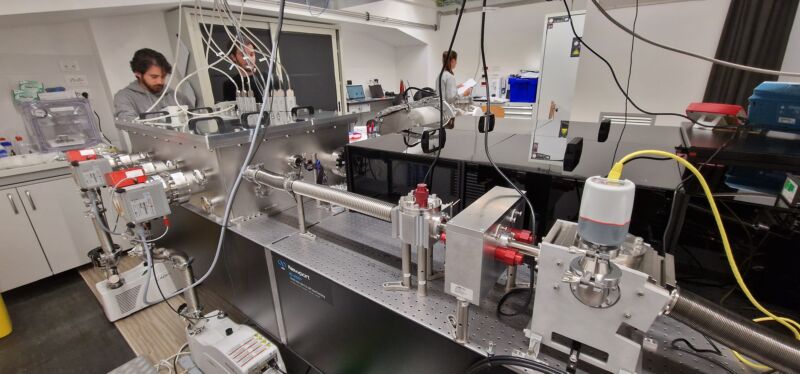
Ultrafast EUV/soft X-ray Microscopy beamline build-up – November 2023
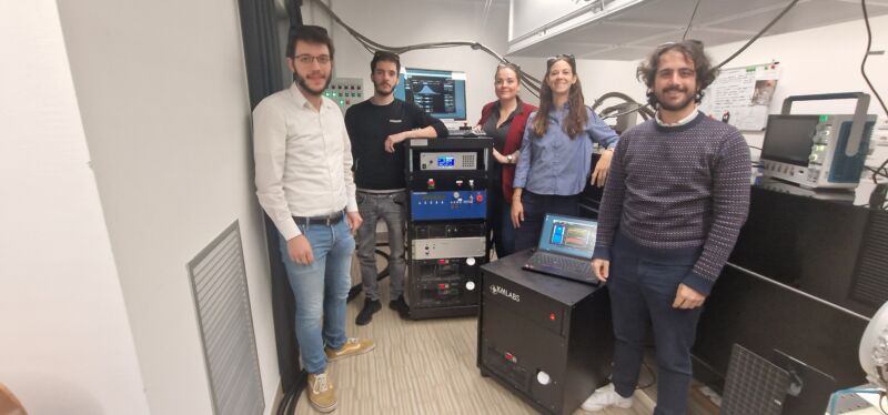
LUXEM group – December 2023
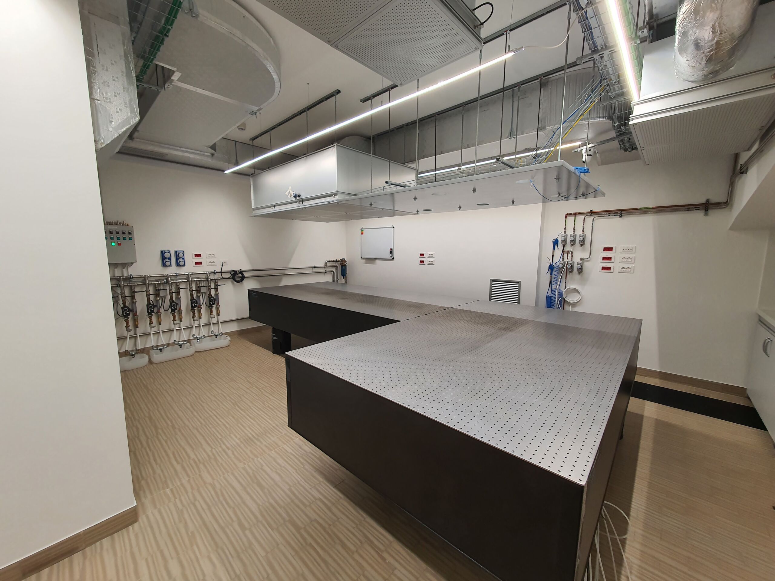
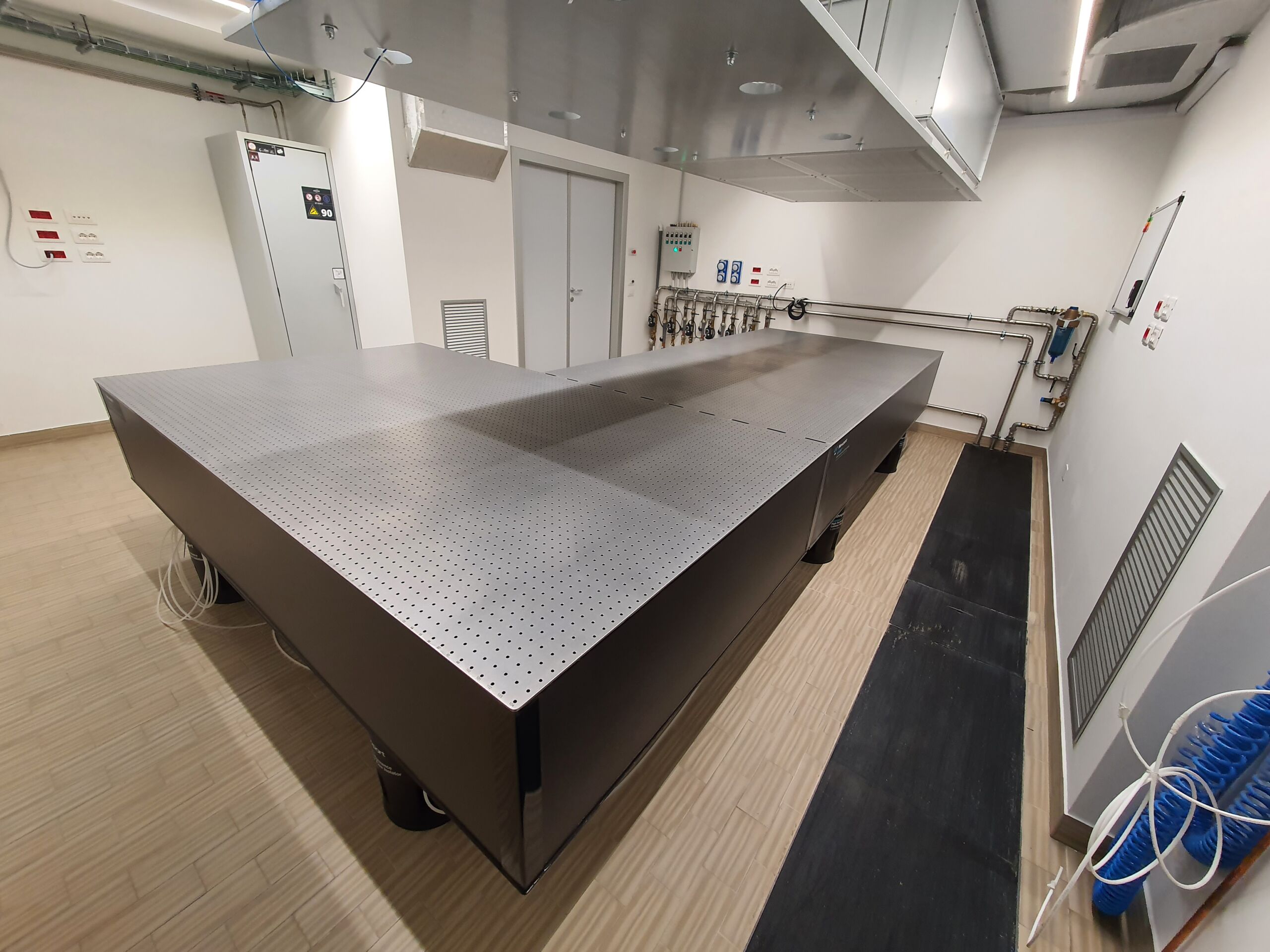
Optical table installation in LUXEM 2 – February 2024
LUXEM 2 infrastructure – February 2024
First non-linear signal from the Ultrafast EUV/soft X-ray Microscopy beamline – May 2024
Optimization of Rhoss UTA performances for LUXEM 1 – July 2024
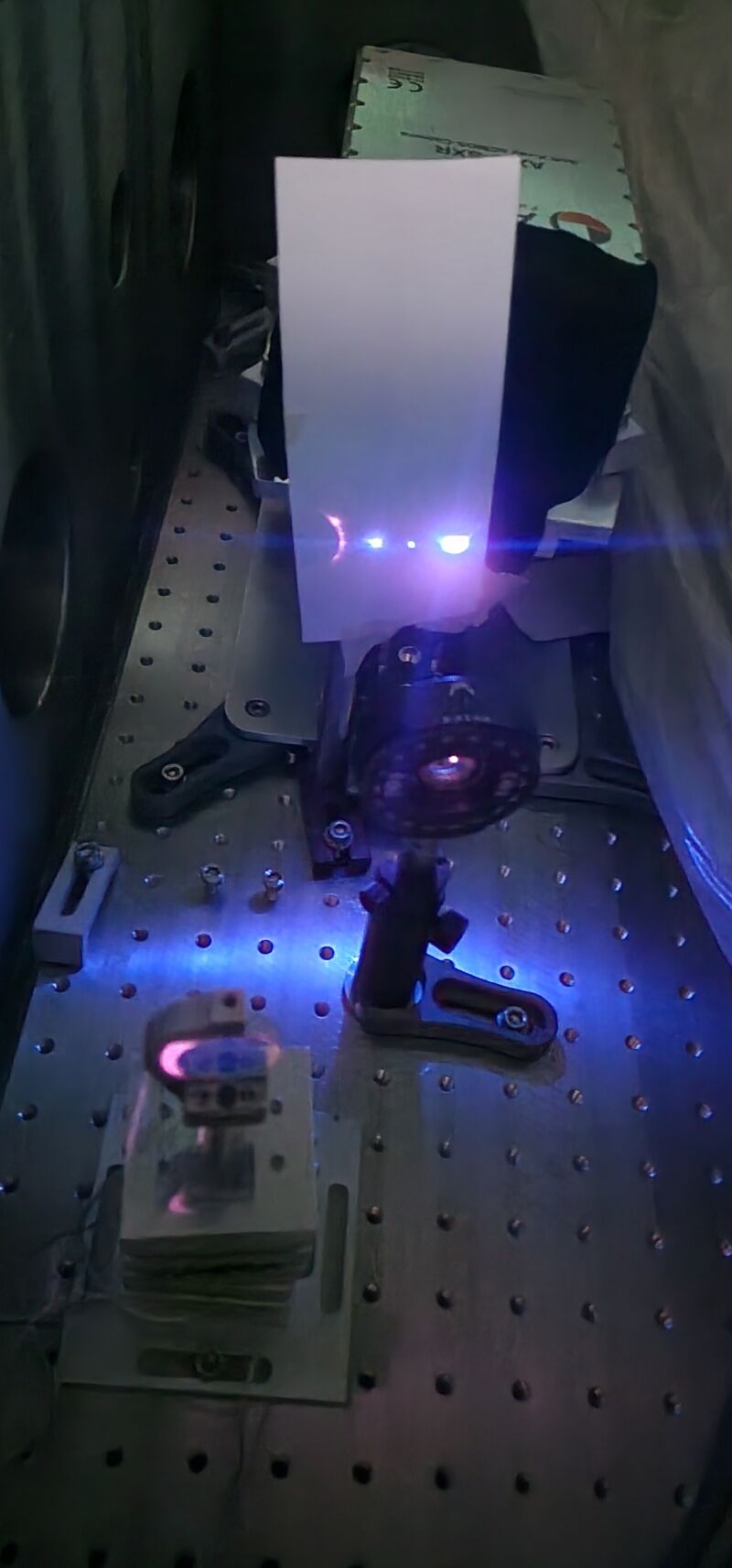
Setting-up for High-Harmonic Generation – September 2024
Preparing the assembly of manipulator and cryostat for the Ultrafast Electron Diffraction and Speckle Imaging beamline – October 2024
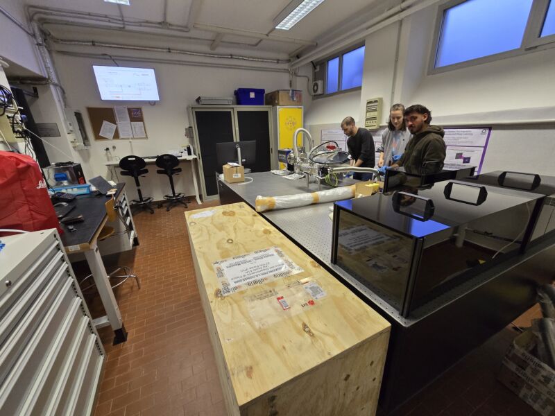
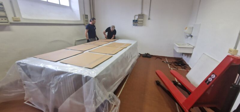
LUXEM 2 infrastructure and prototyping – November 2024
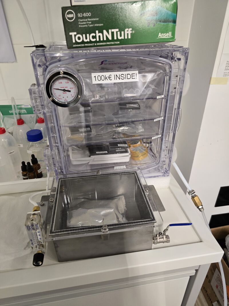
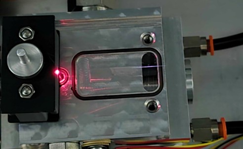
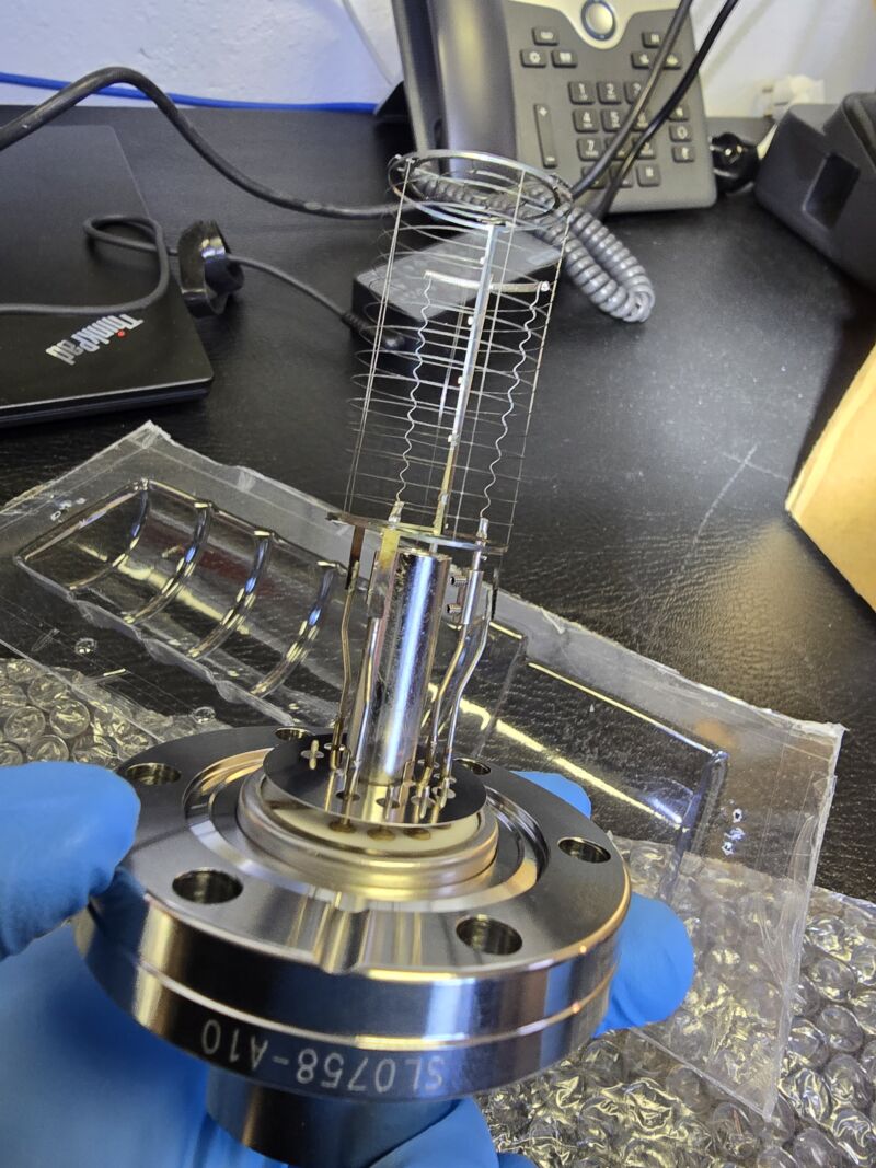
Optics and samples in vacuum – January 2025
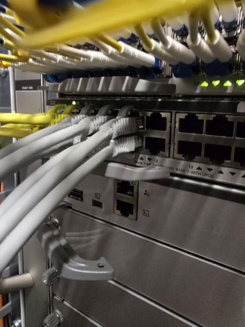
Upgrade of data handling and network across LUXEM cluster, LUXEM 1 and LUXEM 2 – January 2025
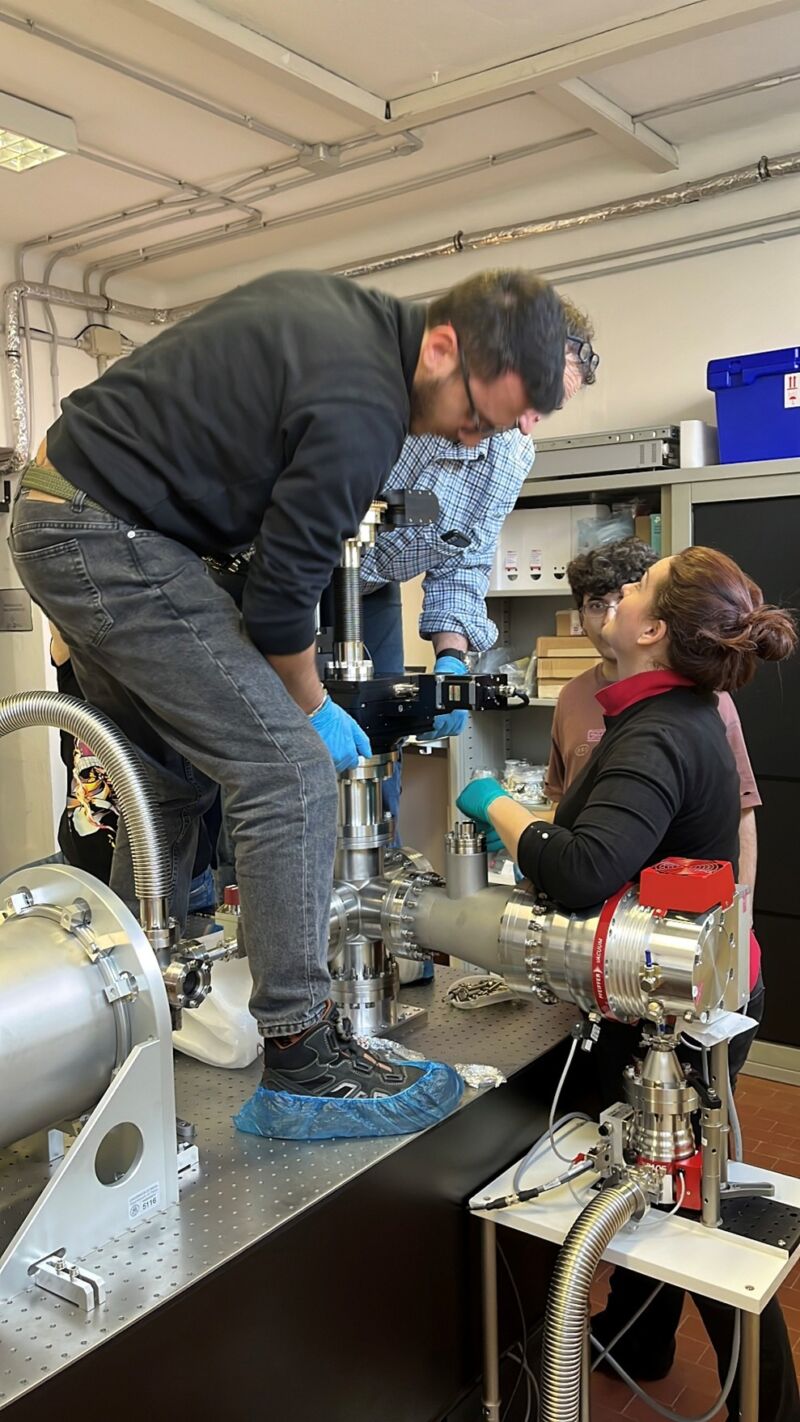
Installation of UED sample manipulator and cryostat – February 2025
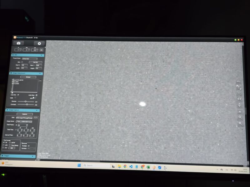
Commissioning completion: Ultrafast EUV/soft X-ray Microscopy beamline – April 2025
UED Ion Gauge troubleshooting completion – June 2025
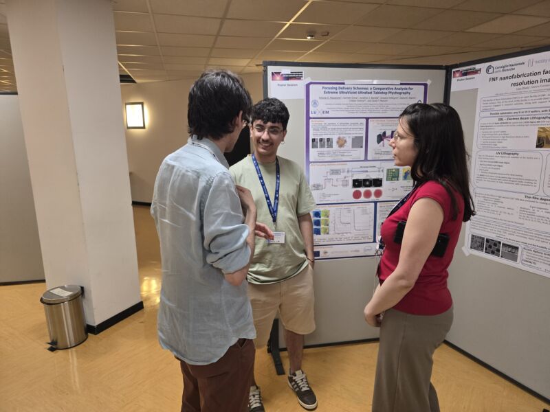
Antonio Mazzarone networking with FERMI FEL peers Francesco Guzzi and Adriana Valerio at AHRI Poster Session – July 2025